4/27/2024
2024 It’s Our Home Sustainability Awards
Celebrating employees whose significant contributions are making a difference, accelerating progress, and creating value while reducing environmental impact.
Read more
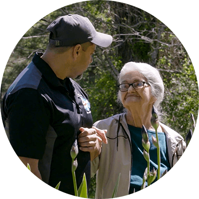
People, Brands and Partners Doing Acts of Good to Help Communities Grow
Discover how P&G and our brands — like Always, Crest, Pampers and Tide — are making a positive impact across communities.
Key P&G Suppliers Honored with 2024 Partner of the Year Awards
P&G Partner of the Year Awards: celebrating suppliers' exceptional contributions and business value.
4/25/2024
Unlocking Solutions to Water Challenges in New “Our Blue World, A Water Odyssey” Documentary
Discover how P&G's partnerships and innovative solutions help address the global water crisis in the documentary "Our Blue World: A Water Odyssey."
Our brands
Align
Probiotic Supplements
Always
Feminine Care Pads
Always Discreet
Incontinence Pads
Ambi Pur
Odor Eliminators
Ariel
Laundry Products
Aussie
Hair Care
Bounce
Dryer Sheets & Fabric Care
Bounty
Paper Towels
Braun
Personal Grooming
Cascade
Dishwasher Detergent
Charlie Banana
Baby Diapers
Charmin
Toilet Paper
Cheer
Clearblue
Pregnancy & Ovulation Tests
Crest
Dental Care
Dawn
Dishwashing Liquid
Downy
Fabric Protectors & Softeners
Dreft
Baby Detergent & Laundry Products
Era
Febreze
Fixodent
Denture Adhesives
Gain
Laundry & Home Products
Gillette
Razors & Skin Care
Head & Shoulders
Herbal Essences
Ivory
Mildly Scented Soap
joy+glee
Razors, Waxes, & Creams
Just
Feminine Care Pads and Tampons
Luvs
Meta
Daily Fiber Supplements
Microban 24
Home Cleaning Products
Mr. Clean
All-Purpose Home Cleaners
Native
Face & Skin Care
Ninjamas
Nighttime Underwear
Olay
Old Spice
Hair & Skin Care
Oral-B
Toothbrushes & Dental Floss
Pampers
Pantene
Pepto-Bismol
Upset Stomach Relief
Prilosec OTC
Heartburn Relief
Puffs
Facial Tissues
Rindex 3en1
Safeguard
Germ-Protecting Soap
Salvo
Scope
Mouthwash
Secret
Deodorant & Body Spray
SK-II
Anti-Aging Face Care
Swiffer
Multi-Surface Dusters & Cleaners
Tampax
Feminine Care Tampons
The Art of Shaving
This is L
Period & Bladder Care
Tide
Venus
Razors & Shaving Gels
Vicks
Cough, Cold & Flu Relief
Zevo
Insect Repellent
ZzzQuil
Sleep Aid
Careers at P&G
Investor Relations